


Chitozen is a chitosan-coated coverslip immobilizing bacteria for microscopy without inducing bacteriostatic effects. It is compatible with 6-channel sticky slides for flow experiments. It is very helpful if you want to:
- Image bacteria both still and alive under the microscope
- Maintain bacteria in a same focal plane for imaging while preserving their physiology
- Renew culture medium, change growth condition (e.g. antibiotics, chemicals, inhibitors) during the experiment and directly observe, in real-time, the bacteria new comportment under the microscope
Sizes:
- 1 slide and 1 sticky slide per pack (TMI-CHI-1)
- 5 slides only (TMI-CHI-7525-min)
- 5 slides and 5 sticky slides per pack (TMI-CHI-7525)
- 1 slide, 1 sticky slide and 4 elements for centrifugation kit (TMI-CHI-1-CENTRI)
- 5 slides, 5 sticky slides and 4 elements for centrifugation kit (TMI-CHI-FULLPACK) Contact us for this product
Catalogue Numbers:
- TMI-CHI-1 (1 slide and 1 sticky slide per pack)
- TMI-CHI-7525-min (5 slides only)
- TMI-CHI-7525 (5 slides and 5 sticky slides per pack )
- TMI-CHI-1-CENTRI (1 slide, 1 sticky slide and 4 elements for centrifugation kit )
- TMI-CHI-FULLPACK (5 slides, 5 sticky slides and 4 elements for centrifugation kit) Contact us for this product
Applications
-
Assay compatibility:
> Fluorescence
> Co-cultures (bacterial predators, immune cells)
> Addition of external factors (e.g. antibiotics, chemicals, inhibitors)
> Static or dynamic conditions -
Imaging modes:
> phase-contrast,
> epifluorescence,
> confocal,
> super-resolution microscopy,
> atomic force microscopy (AFM) - documentation & example pictures available on request -
Experimental outputs:
> Behavioural changes: growth, elongation, cell division, fitness, colony/biofilm formation, etc
> Single molecule imaging -
Chitozen is efficient with the following bacteria:
> E. coli
> Bacillus subtilis
> Caulobacter crescentus
> Corynebacterium glutamicum
> Helicobacter pylori
> Mycobacterium smegmatis
> Myxococcus xanthus
> Pseudomonas aeruginosa
> Pseudomonas fluorescens
> Salmonella
> Staphylococcus aureus
> Vibrio cholerae
This list is updated regularly according to feedback provided by researchers who use Chitozen.
Kit contents
Chitozen coverslips are compatible with 6-channel sticky slides to work in a closed system in static or dynamic flow conditions. Order you coverslips alone if you need to work in an open system (i.e. for AFM imaging).
- 5-coverslips alone*
> 5x standard (25 x 75 mm) chitosan-coated coverslips - 5-coverslips with microfluidic channels (30 experiments)
> 5x standard (25 x 75 mm) chitosan-coated coverslips
> 5x bottomless 6-channel sticky slides
- (Optional) centrifugation pack: the Chitozen coverslips can be centrifuged using a rack compatible with standard microscope slide (25mmx75mm) dimensions. If needed, add our centrifugation pack to your Chitozen kit. It includes:
> µ-Slide Microscopy Rack (ibidi
> Magnetic Lid for Microscopy Rack
> Clamp & adapter for sticky slides
*If using the Chitozen coverslips alone, separate wells can be created manually by using a sealing glue or Stencell silicon chambers. We can provide these 2 products for you. Contact us for more information.
For custom orders (i.e. alternative coverslip amounts), please contact us.
Lifetime: up to 12 months at room temperature, shielded from direct sunlight
Additional resources:> Product overview
> Technical Datasheet
> Safety Datasheet
> FAQs
> Protocol
Results:
Growth and division of E. coli on Chitozen, in LB medium
Credit: Amandine Desorme, LCB - CNRS, 2021
Visualization of Pal mCherry at septum in E. coli (W3110 Pal mCherry), by 3D SIM microscopy, in M9 medium and using Chitozen.
Credit: Amandine Desorme, LCB - CNRS, 2021
Observation of vesicles at septum during cell division of mutant E. coli (W3110 tolR - Palmcherry),
in LB 1/2 medium, using Chitozen.
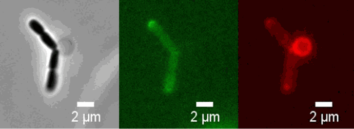
Peptidoglycan is labeled with the green fluorophore BADA.
Credit: Amandine Desorme, LCB - CNRS, 2021
Live cell imaging of E. coli in response to drug addition on Chitozen slides
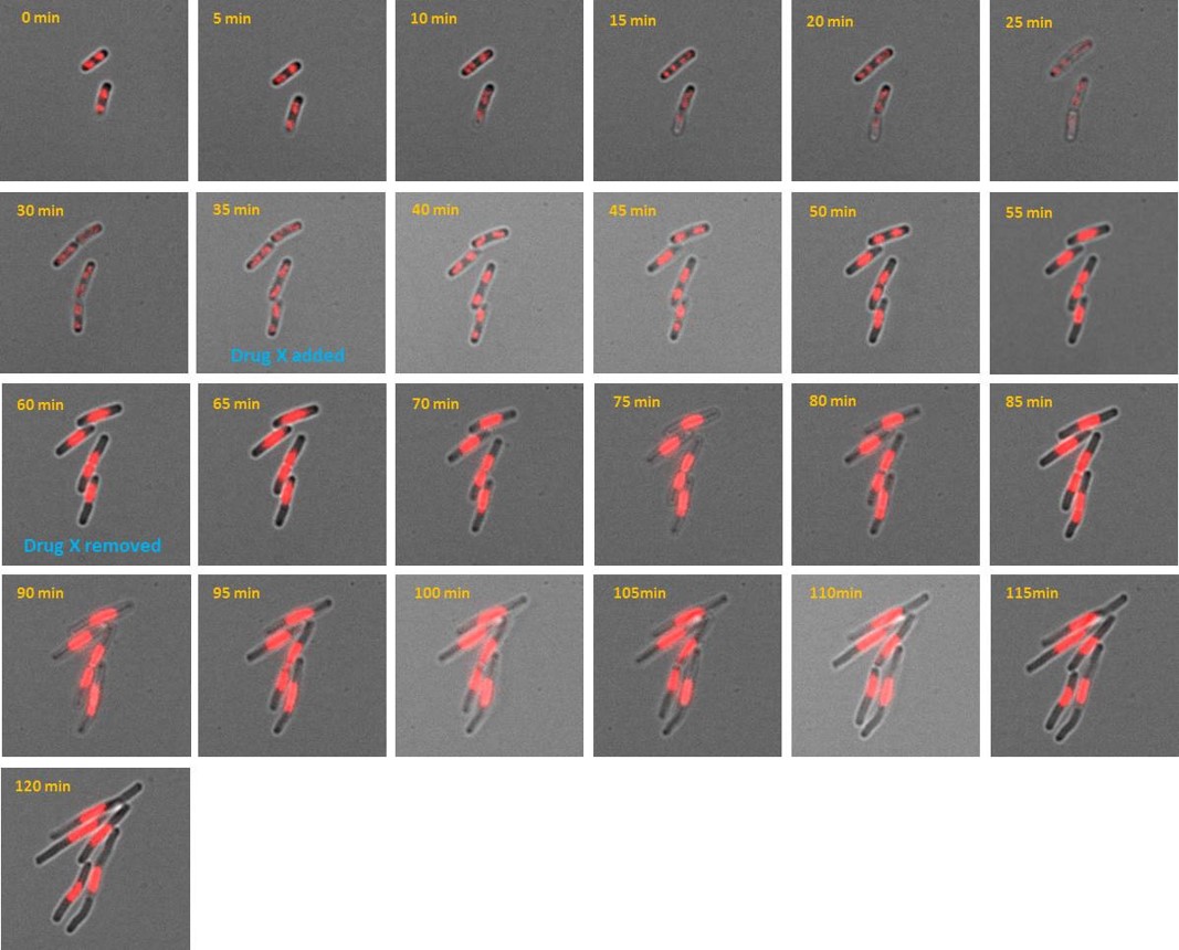
AB1157 E. coli expressing DNA marker HU-mCherry were imaged at 37oC with perfusion of ½ LB at a flow rate of 2 ml/min. Drug X was added at time point 35 minutes and removed at time point 60 minutes.
Credit: Emily Helgesen - Oslo University Hospital - 2022
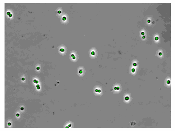
Live cell imaging of E. coli expressing a protein associated with DNA replication on Chitozen slides
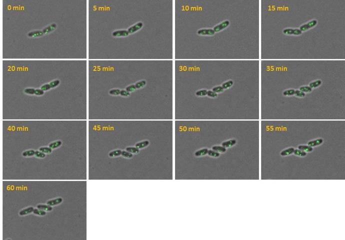
AB1157 E. coli expressing SeqA-YFP (green), a protein associated with DNA replication, were imaged at 37°C over 60 minutes without perfusion of medium
Credit: Emily Helgesen - Oslo University Hospital - 2022
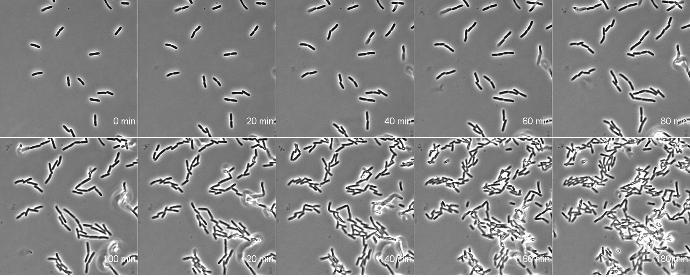
Effects of cell division inhibitor cephalexin on E. coli growth cultured on Chitozen slides
E. coli BW25113 cells were imaged at 37°C with perfusion of M9 medium at a flow rate of 0.05 mL/min.
Credit: Bianca Sclavi - 2021
E. Coli spheroblasts bound to Chitozen
E. coli MG1655 with chromosomally encoded HU-eGFP under the native promoter were imaged with perfusion at a flow rate of 0.05 mL/min.
Credit: Itzhak Fishov – Ben-Gurion University of the Negev, 2022